Structure And Function Of Respiratory Organs
Respiration is the process of exchanging respiratory gases between the external environment and the body. This essential process involves the oxidation of food and the liberation of energy. Respiration takes place in a specialized organ system known as the respiratory system. #Respiratory Organs
PHYSIOLOGY OF RESPIRATIONPHYSIOLOGY
Structure and Function of the Respiratory Organ
Respiration: An Overview
Respiration is the process of exchanging respiratory gases between the external environment and the body. This essential process involves the oxidation of food and the liberation of energy, which is necessary to maintain various life activities. Respiration takes place in a specialized organ system known as the respiratory system.
Features of the Respiratory System
The respiratory system is responsible for the exchange of respiratory gases due to several key features:
1. Thin Membrane: The respiratory system has a thin membrane that facilitates efficient gas exchange.
2. Increased Surface Area: The system has an increased surface area to maximize the exchange of gases.
3. Respiratory Epithelium: The respiratory epithelium and the endothelium of blood vessels are closely associated, allowing for effective gas exchange.
4. Vascular Nature: The respiratory system is highly vascular, meaning it has a rich supply of blood vessels. Its lining is made up of involuted cells, which further aids in the respiratory process.
Division of the Respiratory System
The respiratory system of mammals, including humans, is divided into two distinct regions:
The Respiratory Route
The Respiratory Seat (or lobe)
Respiratory Route
The respiratory route consists of several organs responsible for the transport of respiratory gases. These include:
1. External Nares (Nostrils):
These are the paired openings present at the tip of the nose.
2. Nasal Chambers:
The nasal cavity is divided into two chambers by a vertical partition called the nasal septum. Internally, each nasal chamber can be divided into three regions:
- Vestibular Region: This is the outermost part of the nasal chambers, lined by stratified epithelium containing a few sebaceous glands and hair. This region acts as a sieve to filter incoming air.
- Respiratory Region: This is the middle region of the nasal chambers, lined by pseudostratified columnar epithelium. This region plays a significant role in warming and humidifying the air.
- Posterior Olfactory Region: The posterior olfactory region is lined with olfactory epithelium, which functions as the organ of smell.
3.Internal Nares:
The nasal chambers open internally into the pharynx through an opening called the internal nares. During swallowing, the internal nares get closed by the soft palate to prevent food from entering the nasal cavity.
4.Pharynx
The pharynx is a short muscular chamber located behind the soft palate and extending to the gullet and glottis. It is divided into three functional regions:
1. Nasopharynx: The upper part connected to the nasal cavity.
2. Oropharynx: The middle part behind the oral cavity.
3. Laryngopharynx: The lower part leading to the larynx and esophagus.
5.Glottis and Epiglottis
The glottis is a slit-like opening on the floor of the laryngopharynx.
It is covered by a flap-like cartilage called the epiglottis, which closes the glottis during the passage of food to prevent it from entering the respiratory tract.
6.Larynx
The glottis leads into the larynx, a cartilaginous and ciliated chamber located in front of the laryngopharynx. In adult men, the larynx grows larger and becomes prominent on the front of the neck, forming the Adam's apple. The larynx continues downward and opens into the trachea. In mammals, the larynx functions as the sound-producing organ.
7.Trachea
The trachea, or windpipe, is a long tube about 19 cm long and 2.5 cm wide in adult humans.
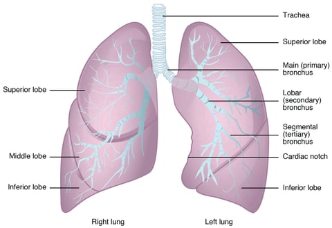
It runs down beneath the esophagus in the neck region and enters the thoracic cavity. At the level of the 5th and 6th ribs, it divides into a pair of primary bronchi, each entering one lung. The walls of the trachea are reinforced by C-shaped rings of cartilage (16-20), providing flexibility and protection against collapsing.
Internally, the trachea is lined with pseudostratified ciliated columnar epithelium containing mucus-secreting goblet cells, which keep the trachea moist.
8.Bronchi
The trachea divides into two primary bronchi, which further divide into secondary bronchi. Each secondary bronchus divides into tertiary bronchi. The inner walls of all these bronchi are lined with ciliated mucosa and supported by incomplete cartilaginous rings.
9.Lungs
The lungs act as the primary respiratory organs in mammals and other vertebrates.
They are located in the thoracic cavity, supported dorsally by the vertebral column, ventrally by the sternum, and laterally by the ribs.
The thoracic diaphragm supports the lungs from below.
The space between the lungs is called the mediastinum.
Each lung is lodged in the pleural cavity, lined by an inner visceral pleura and an outer parietal pleura.
The fluid in the pleural cavity acts as a lubricant, preventing wear and tear and providing protection against mechanical shock.
Each lung has a spongy and elastic structure with a pinkish color and conical shape.
The top part is called the apex, and the broader bottom part is the base. The left lung has two lobes, while the right lung has three lobes.
Internal Structure of the Lung Lobes
Each lobe of the lung is internally divided into lobules.
A lobule receives a terminal bronchiole.
The terminal bronchiole is further divided into respiratory bronchioles. The structure of the respiratory bronchiole is intricate and folded. Each respiratory bronchiole is again divided into alveolar ducts.
The alveolar ducts expand to form the atria. The atria lead into alveolar sacs, each of which contains multiple small, flask-shaped pouches called alveoli. There are approximately 750 million alveoli in total, covering a surface area of about 100 square meters, which is 100 times greater than the surface area of human skin.
The alveoli contain lipoprotein in their membranes, which prevents the walls from collapsing during expiration. The lining of the alveoli is formed of squamous epithelium, which is provided with a network of blood capillaries on the outer surfaces, along with some reticular and elastic connective tissue.
The squamous epithelium of the alveoli contains cuboidal cells that secrete a thin film of surfactant, which is a lipoprotein. This surfactant reduces surface tension and prevents the alveoli from collapsing.
Engage with Us:
Stay tuned for more captivating insights and News. Visit our Blogs and Follow Us on social media to never miss an update. Together, let's unravel the mysteries of the natural world.


