
Fate Mapping: Method & Techniques Notes
Explore the concept of fate mapping, including its history, techniques like natural markers, and real-world examples. Understand how scientists use fate maps to track embryonic development in various organisms.
STUDY ZONE
Fate Map
A fate map is a diagrammatic representation of the prospective fate of each part of an embryo at an early stage of development.
The different methods used for constructing fate maps are as follows:
1. Natural Markers
Fertilized eggs of ascidians, such as Ciona and Styela partita (Cynthia), exhibit natural color differences in various regions of the egg. In such cases, it is possible to trace the fate of each blastomere.
E.G. Conklin, while studying the fate of each blastomere, observed that:
Cells containing clear cytoplasm formed ectoderm.
Dark grey yolky cells formed endoderm.
Light grey cells gave rise to the notochord and neural plate.
Cells with yellow cytoplasm developed into mesoderm.
However, in many organisms where natural color differentiation does not occur, artificial methods must be employed.
2. Artificial Markers
Fate maps can be constructed by labeling single cells or specific regions of the embryo and then tracking the position and shape of the labeled patch.
Vital Dyes: Vogt (1925) used vital dyes to stain early developmental stages, such as the blastula.
The dyes used include:
Nile blue sulfate
Neutral red
Bismarck brown
These dyes do not interfere with cell function but help trace the fate of cells.
The staining procedure involves are given below:
1. Mixing the dye with agar and spreading it on a slide to dry.
2. Pressing the dried agar piece against a chosen area of the blastula for a short duration.
3. Allowing the stain to diffuse from the agar into the blastomeres.
Note:
This method enables the simultaneous marking of multiple areas of the embryo, allowing researchers to track their fate during development.
Carbon Particles: N. Spratt (1946) marked blastomeres with carbon particles to construct the fate map of a chick embryo. These particles adhered to the cell surface, allowing researchers to trace cell fate. Later, William W. Ballard (1981) modified this technique by injecting carbon or chalk particles into specific regions of teleost embryos to track their developmental fate.
Radioactive Markers: Radioactive markers such as C¹⁴, P³², and H³ have been used to study cell lineage. Tritiated thymidine labels the nuclei as it integrates into the DNA of dividing cells.
The fate of these labeled cells is traced using autoradiography, which involves:
1. Cutting a specific region from the host embryo.
2. Replacing it with a radioactive graft from a donor embryo.
3. Tracking the descendants of the labeled cells, which appear distinct from unlabeled cells under autoradiographic analysis.
Note:
However, vital dyes and radioactive labeling pose challenges in fate map construction, as they become diluted with each cell division.
Fluorescent Dyes: To overcome the limitations of vital dyes and radioactive markers, fluorescent dyes have been introduced. Fluorescein-conjugated dextran can be injected into cells, and its progeny can be easily detected. Common fluorescent dyes include:
Fluorescein Dextran Amine (FDA)
Rhodamine Dextran Amine (RDA)
Additionally, carbocyanine dyes such as DiI (red emission) and DiO (green emission) are lipophilic membrane stains that diffuse through the cell and can be visualized under a fluorescence microscope.
Histochemical Stains: This method relies on enzyme-specific staining of embryonic cells. One commonly used stain is horseradish peroxidase (HRP), which becomes visible when an appropriate substrate is added to trigger enzymatic activity.
3. Genetic Markers
Compared to vital dyes and other stains, genetic markers offer the advantage of not spreading to neighboring cells and being inherited by the progeny of marked cells if stably expressed. However, the low efficiency of gene introduction into cells remains a challenge.
Chimeric Embryos
Genetic markers are used to create chimeric embryos by transplanting embryonic tissues from organisms with different genetic constitutions but similar developmental patterns. This process, called xenoplastic transplantation, has been demonstrated using quail and chick embryos, which differ in their heterochromatin positioning and cell surface antigens.
Retrovirus Marking
Retrovirus marking is another technique used to study cell fate by incorporating a retrovirus-engineered reporter gene into the DNA of host cells. Once expressed, the reporter gene’s product can be detected using histochemical or fluorescent methods.
Retroviruses are RNA viruses that contain reverse transcriptase, an enzyme that enables them to create a DNA copy of their genome upon infecting a host cell.
This viral DNA integrates into the host cell’s chromosome and is passed on to progeny cells, forming transgenic DNA chimeras.
Additionally, host DNA can be genetically modified to express green fluorescent protein (GFP), a naturally occurring protein in some jellyfish, which emits a green fluorescence under appropriate lighting conditions.
Genetic Labeling – Brainbow Technique
More recently, advance genetic labeling techniques such as the Brainbow method have been employed for lineage studies. This technique involves tagging different proteins in the cells with fluorescent proteins, generating a spectrum of colors through distinct fluorescent protein expression. The array of colors allows researchers to distinguish neurons in the brain, hence the name Brainbow. Since its introduction, this method has been used to trace the fate of early embryonic structures during development and to enhance our understanding of fate maps.
ADVERTISEMENTS
ADVERTISEMENTS
Significance of Fate Maps
Fate maps are essential tools for tracing cell lineage, allowing scientists to follow the development of specific embryonic regions.
1. Animal Pole Development: The animal pole of the egg gives rise to the future ectoderm, which further differentiates into :-
Prospective non-neural ectoderm – develops into the epidermis of the skin.
Prospective central nervous system – gives rise to the brain, spinal cord, and sense organs.
2. Marginal Region Development: The marginal region of the gray crescent develops into presumptive mesodermal cells, which are divided into the following subregions:
Presumptive notochordal region – gives rise to the notochord.
Foregut region – lies below the notochordal area.
Presumptive somites – develop on both sides of the notochordal area.
Ventrolateral mesodermal area – found in the lateral and ventral parts of the marginal zone, forming:
The mesodermal lining of the body cavity.
Kidneys
Reproductive organs
Extraembryonic membranes
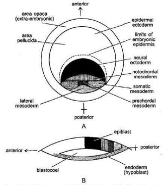
Fate Map of a Chick Embryo
The fate map of the chick epiblast reveals the following key regions:
1. The anterior half forms the prospective epidermis and extraembryonic membranes.
2. Below the epidermis lies the presumptive neural tissue.
3. Behind the neural tissue is the presumptive notochord region.
4. The presumptive mesoderm is located along the midline behind the notochord, with most of the posterior half of the area pellucida forming the mesodermal lining of the body cavity.
Fate Map of Mammals
Constructing a fate map for human blastula is particularly challenging due to intrauterine development. However, fate maps have been prepared for other mammals such as mice, rabbits, and monkeys.
The epidermal ectoderm gives rise to neural tissue, epidermis, and notochord.
The hypoblast contributes to the formation of endodermal structures.
These fate maps serve as critical references for understanding mammalian embryonic development.

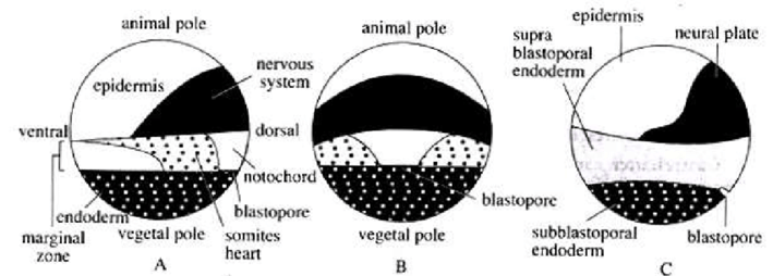

Fate Map of Frog Egg: Late Blastula (A); Dorsal View (B); Exterior View (c) Showing Presumptive Organ forming Areas
Fate Maps of Chick Blastoderm: Surface View (A); Section of Discoblastula
References
1. Gilbert, S. F. (2019). Developmental biology (11th ed.). Sinauer Associates.
2. Helmchen, F., & Konnerth, A. (Eds.). (2011). Imaging in neuroscience: A laboratory manual. Cold Spring Harbor Laboratory Press.
3. Bear, M. F., Connors, B. W., & Paradiso, M. A. (2020). Neuroscience: Exploring the brain (4th ed.). Jones & Bartlett Learning.
4. Kimmel, G. L., & Kochhar, D. M. (Eds.). (1990). Molecular and cellular methods in developmental toxicology. CRC Press.
5. Kandel, E. R., Schwartz, J. H., & Jessell, T. M. (Eds.). (2013). Principles of neural science (5th ed.). McGraw-Hill.
ADVERTISEMENTS
ADVERTISEMENTS
ADVERTISEMENTS
ADVERTISEMENTS

Drop Us a Line
We’d Love to Hear from You