Nature of the Curve:-
🦨The curve shows a steep rise in % saturation of hemoglobin (Hb) between O₂ tensions of 0 to 40 mmHg, indicating a significant increase in oxygen binding.
🦨There is no notable increase in the % saturation of Hb between O₂ tensions of 40 to 60 mmHg.
🦨The graph indicates that between O₂ tensions of 60 to 100 mmHg, there is no significant increase in the % saturation of Hb.
🦨The first part of the curve is nearly vertical, while the second and third parts of the curve are almost parallel to the X-axis.
🦨This graph indicates that the increase in % saturation of Hb is due to the increase in partial pressure of O₂ (Po₂). When there is a decrease in O₂ tension, O₂ is dissociated at the site of tissues because of low partial pressure of oxygen and high CO₂ levels.
Factors Affecting the Dissociation Curve:-
🦨The dissociation curve is influenced by several factors, which affect the binding and release of oxygen by hemoglobin.
🦨These factors are crucial in understanding the physiological adaptations of various organisms.
1.Temperature:
🪶An increase in temperature decreases the saturation of hemoglobin with oxygen.
🪶Example: At 38°C and a partial pressure of oxygen (Po₂) of 100 mm Hg, the saturation of hemoglobin might drop significantly. Conversely, at 25°C, hemoglobin saturation is much higher.
🪶Warm-blooded animals dissociate oxygen more efficiently and rapidly than cold-blooded animals.
2.Electrolytes:
🪶At low oxygen tension, oxyhemoglobin releases more oxygen in the presence of electrolytes compared to a pure solution.
🪶The dissociation of oxyhemoglobin is facilitated by an increase in hydrogen ions (H+), which occurs due to the presence of CO₂ in the blood, leading to increased acidity.
3.pH and CO₂ Effect:
🪶The Bohr Effect describes how an increase in CO₂ and a decrease in pH cause a rightward shift in the oxygen dissociation curve.
🪶During intense muscular contraction, CO₂ and acidic products increase, leading to a decrease in pH. This decreases hemoglobin’s affinity for oxygen, causing more oxygen to be released to tissues.
🪶In environments with high CO₂, the formation of oxyhemoglobin is reduced, leading to insufficient oxygen supply for energy production. This results in increased breathing rates and a feeling of breathlessness.
4.Effect of pH:
🪶The binding capacity of hemoglobin is decreased by altering the pH.
🪶This effect, observed by Bohr, indicates that changes in pH can be either normal or excessive, depending on the rise or fall in pH.
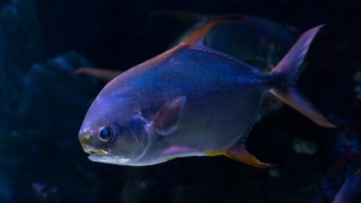
🪶The Root Effect, noted in many fish species, illustrates that at lower pH levels, less oxygen is carried by hemoglobin.
🪶These factors collectively influence how efficiently oxygen is transported and utilized in the body, highlighting the adaptability of different organisms to their environments.
Carbon Monoxide (CO) Poisoning
🪶A condition caused by inhaling carbon monoxide (CO).
🪶Incomplete combustion of fuels like gasoline, propane, and natural gas in appliances or vehicles.
🪶CO is colorless, odorless, and tasteless, making it difficult to detect.
🪶CO binds to hemoglobin in red blood cells more readily than oxygen, depriving the body of oxygen.
🪶Infants and unborn babies,elderly people and Individuals with chronic health conditions like heart disease, anemia, or respiratory problems.
🪶During colder months when furnaces and heaters are in use.
Mechanism of CO poisoning:-
* CO enters the bloodstream through the lungs.
* Hemoglobin in red blood cells preferentially binds to CO over oxygen.
🪶Oxygen Binding to Myoglobin:
Oxygen (O₂) binds with myoglobin via a nitrogen atom of the heme group.
💫Myoglobin-Oxygen Complex: Nb+ O2–>NbO2
🪶Hemoglobin Binding:
Hemoglobin (Hb) can bind oxygen in a similar manner, forming HbO2.
🪶The dissociation constant will determine the strength of the carbon monoxide (CO) bond.
🪶A higher dissociation constant indicates weaker bonding.a lower dissociation constant means stronger bonding.
Respiratory Pigments
🪶In order to facilitate the transport of oxygen to different parts of the body, most animals have developed respiratory pigments.
🪶Respiratory pigments are of profound physiological importance, especially in large-sized animals, because uniform distribution to all parts of the body by simple diffusion would be difficult.
🪶Types of Respiratory Pigments;Four different respiratory pigments are recognized:
– Haemoglobin
– Chlorocruorin
– Hemocyanin
– Hemerythrin
🪶Even in the same phylum, there may be several distinct pigments.
🪶More than one distribution of respiratory pigments in the same animal may exist.
Haemoglobin
🪶Haemoglobin is the most efficient and widely distributed respiratory pigment in the animal kingdom. It is found in:
Some protozoa like Paramecium.
🪶Almost all vertebrates except eel larvae and some Antarctic fishes.
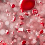
🪶In vertebrates, haemoglobin is contained inside erythrocytes (red blood cells).
🪶In invertebrates, it may remain dissolved in the blood plasma or be contained in special cells called erythrocytes.
Structures of Haemoglobin
🪶Vertebrate hemoglobin has molecular weights varying between 64,000 and 68,000 Daltons.
🪶Invertebrate hemoglobin generally has low molecular weights, ranging between 15,000 Daltons to 1-3 million Daltons.
🪶Structure of Hemoglobin:
Hemoglobin is made up of a non-protein compound called heme, associated with a protein called globin.
💫Heme is composed of 4 pyrrole rings linked by methine bridges (-CH=) to form a porphyrin ring, with a ferrous ion (Fe²⁺) in the center.
🪶The heme component is a constant structural feature of all hemoglobins, but the globin portion varies in different species.
🪶Variation in Hemoglobin:
In addition, the number of hemoglobin polymers varies, leading to different forms of hemoglobin across species.
🪶In adult humans, 90 percent of hemoglobin consists of two alpha chains combined with two beta chains, forming hemoglobin A. Each alpha chain contains 141 amino acids, while each beta chain contains 146 amino acids.
🪶Adult blood also contains a small amount of hemoglobin A2, in which the beta chains are replaced by delta chains, and a minor fraction of hemoglobin F, which has gamma chains.
🪶Hemoglobin binds to oxygen through its ferrous iron atoms, each of which can associate with one molecule of oxygen to form oxyhemoglobin.
🪶This binding reaction is readily reversible, allowing hemoglobin to release oxygen where it is needed. Oxyhemoglobin is red in color both in its oxygenated and deoxygenated forms.
🪶Oxygen Saturation:
The association of hemoglobin with oxygen depends on the pH and ionic content of the blood.
🪶The partial pressure of oxygen at which hemoglobin is half-saturated with oxygen varies among different organisms:
– 10-20 mmHg in fish
– 20-40 mmHg in land vertebrates
– 40-60 mmHg in birds
– Generally below 10 mmHg in invertebrates Haemocyanin
1.Nature and Composition:
👉Haemocyanin is a copper-containing protein found in nature.
👉 It occurs in certain arthropods and mollusks, serving as a respiratory pigment.
2.Dissolution in Plasma:
👉Haemocyanin always remains dissolved in plasma.
👉The molecular weight is very high, ranging from 400,000 Daltons to 13,000,000 Daltons in gastropods.
3.Structure and Subunits:
👉The molecules of haemocyanin are larger than those of hemoglobin.
👉Haemocyanin consists of a greater number of subunits compared to hemoglobin.
4.Absence of Porphyrin:
👉Haemocyanin does not contain porphyrin.
Structure of Haemocyanin
1.Molecular Structure:
The molecules of haemocyanin, which include copper, are composed of a peptide chain with just over 200 amino acids.
2.Oxidation State:
During oxygenation, the copper atoms in haemocyanin are oxidized to the cupric form.
3.Deoxygenated State:
In deoxygenated haemocyanin, the copper is in the cuprous form. The deoxygenated form is colorless but changes in appearance when oxygenated.
4.Oxygen Binding:
The pigment binds with oxygen at different concentrations depending on various factors.
5.Oxygen Transport Capacity:
The oxygen transport capacity of haemocyanin is less than that of haemoglobin.
Chlorocruorin
1.Color and Location:
This green-colored metalloprotein is found in the plasma of certain polychaete families, such as Sabellidae and Serpulidae.
2.Metal Composition:
It is a metalloprotein with a metal component similar to heme, but the vinyl group (CH-CH2) is replaced by formyl (CHO) groups in chlorocruorin.
3.The porphyrin structure in chlorocruorin is called chlorocruorin, except in some variations.
Hemerythrin
1.Color and Location:
This violet-colored pigment is found inside the corpuscles of marine organisms belonging to the phyla Sipunculidae, Priapulida, and Brachiopoda, and also in the polychaete worm Magelona.
2.Metal Composition: Hemerythrin is a non-heme iron-containing metalloprotein.
3.Function: It is involved in oxygen transport and storage, similar to other respiratory pigments.


✦✦✦✦✦


